Welcome to our Publications Page
Discover a selection of whitepapers and case studies highlighting Tecdia’s innovative solutions across diverse industries. Explore our contributions that have made a real difference to our customers’ success stories. We take pride in the valuable contributions our solutions have made across diverse industries, driving progress and enabling innovation.
Mentions of Tecdia Ceramic Products
Publications (Click for Abstracts/Link)
- The design of power amplifier for 5.8 GHz wireless LAN application using GaAs substrate
- Visual observation of internal signal transmissions in a millimeter-wave amplifier module
- Live electrooptic imaging of K-band switching actions and parasitic phenomena in MMIC module
- A Directly Matched PA-Integrated K-Band Antenna for Efficient mm-Wave High-Power Generation
- E-band filters based on substrate integrated waveguide octagonal cavities loaded by complementary split-ring resonators
- Crystalline Flat Surface Recovered by High-Temperature Annealing after Laser Ablation
- Broadband Device Modeling and Q Calculations of Barium Strontium Titanate (BST) Varactors
- High-Q Millimeter Wave RF Filters and Multiplexers
The design of power amplifier for 5.8 GHz wireless LAN application using GaAs substrate

This paper describes a compact two-stage hybrid power amplifier for 5.8 GHz-band Wireless Local Area Network (WLAN) applications. The amplifier has been fabricated on high dielectric semiconductor wafer, and then the amplifier has small dimension compared with the conventional hybrid circuits. To fabricate the amplifier, the mask pattern is produced using electron beam lithography. Then the FET, capacitors and resistors are combined with the microstrip line pattern by silver epoxy. The size of the two-stage amplifier is measured to be 2.1 cm/spl times/1.4 cm.
Visual observation of internal signal transmissions in a millimeter-wave amplifier module

The internal signal transmissions in a millimeter-wave module were visually observed for the first time. The module contains a 2-35 GHz GaAs travelling wave amplifier chip and microstrip lines on alumina substrates. In the frequency range of 10-35 GHz, visual observation was achieved using a live electrooptic imaging camera based on the optical magnification method. It was successfully demonstrated that the voltage-dependent performance of the amplifier and symptoms of parasitic cavity resonance effect were visualized.
Live electrooptic imaging of K-band switching actions and parasitic phenomena in MMIC module

It has been successfully demonstrated that K-band signal behaviors in a monolithic microwave integrated circuits (MMIC) module can be visually observed and analyzed. The visual observations and analyses were performed experimentally by the live electrooptic imaging technique. The module consists of three radio-frequency ports and a MMIC chip of DC-50 GHz single pole double throw switch. Its basic switching actions were visualized clearly while the following parasitic phenomena were visually captured; cavity resonances, their dependences on the module operation modes, and coupling between the input and output lines.
A Directly Matched PA-Integrated K-Band Antenna for Efficient mm-Wave High-Power Generation

A K-band slot antenna element with an integrated gallium nitride power amplifier (PA) is presented. It has been optimized through a circuit-electromagnetic codesign methodology to directly match the transistor drain output to its optimal load impedance (ℤ opt = 17 + j46 Ω), while accounting for the over-the-air coupling effects in the vicinity of the transition between the PA and antenna. This obviates the need for using a potentially lossy and bandwidth-limiting output impedance-matching network. The measured PA-integrated antenna gain of 15 dBi with a 40% total efficiency at 28 dBm output power agrees well with the theoretically achievable performance targets. The proposed element is compact (0.6 × 0.5 × 0.3 λ 3 ) and, thus, well suited to meet the high-performance demands of future emerging beamforming active antenna array applications.
E-band filters based on substrate integrated waveguide octagonal cavities loaded by complementary split-ring resonators

In this paper, substrate integrated waveguide (SIW) octagonal cavities in conjunction with octagonal complementary split-ring resonators (CSRRs) are used to propose compact narrow bandpass filters at E-band. Short circuited slots are employed as coplanar waveguide (CPW) to SIW transitions as well as matching elements between adjacent stages of filters. Parameterized models with full-wave accuracy are extracted for three different SIW-CSRRs unit cells. Each unit cell is comprised of an SIW cavity, a pair of octagonal CSRRs etched on the top plate, and a CPW to SIW transition with a certain slot length for bandwidth adjustment. These unit cell models are used to design and optimize multi-stage E-band filters having five, six and seven stages. Measured results of filters show insertion loss, ripple and group delay variation on the order of 7-dB, 1-dB and 30 ps over the passband of 70-73 GHz while about 30-dB rejection at 66.5 GHz and 76.5 GHz is achieved.
Crystalline Flat Surface Recovered by High-Temperature Annealing after Laser Ablation
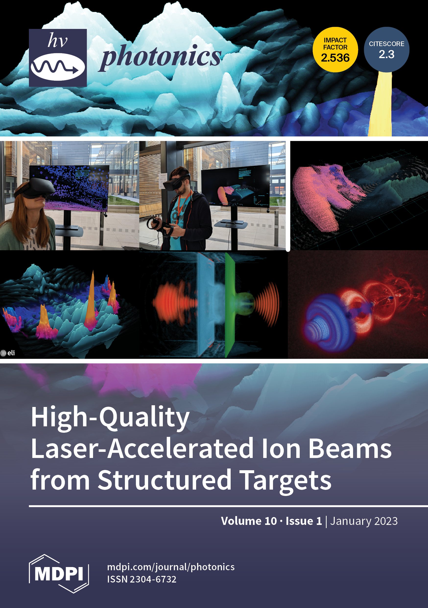
Ultra-short laser pulses (1030 nm/230 fs) were used to laser ablate the surface of crystalline sapphire (Al2O3) at high intensity per pulse 20–200 TW/cm22/pulse. Laser-ablated patterns were annealed at a high temperature of 1500 ∘°C. Surface reconstruction took place, removing the ablation debris field at the edges of ablated pits in oxygen flow (O2 flow). Partial reconstruction of ripples was also observed when multi-pulse ablated surfaces were annealed at high temperature in O2 flow. Back-side ablation of a 0.5-mm-thick Al2O3 produced high surface roughness ∼1μ1 m which was reduced to ∼0.2μ0.2 m by high-temperature annealing at 1500 ∘°C for 2 h in O2. Improvement of surface quality was due to restructuring of the crystalline surface and sublimation, while the defined 3D shape of a micro-lens was not altered after HTA (no thermal morphing).
Broadband Device Modeling and Q Calculations of Barium Strontium Titanate (BST) Varactors

Varactors are used in a wide variety of RF and Microwave circuits. Manufacturers of these devices typically offer electrical models which Design Engineers can use to include in their design simulations to achieve first-pass design success. This paper describes the measurement, electrical model optimization and Q calculations of Barium Strontium Titanate (BST) varactors. The measurement and modeling process described can be used as a template to model any varactor technology.
High-Q Millimeter Wave RF Filters and Multiplexers

For a long period of time, millimeter waves (mm-Wave) were considered unsuitable for wireless data transmission due to high attention while propagating in the atmosphere. Over the past few years, due to the vigorous developments of multiple-in-multiple-out (MIMO) antenna technology and semiconductor technology, it is now feasible to have reliable wireless data transmissions using mm-Wave. Traditionally, mobile communication networks operate in the frequency spectrum under 6 GHz. In order to meet the ever-increasing demand for high communication data rate and high-quality multi-media services, the current fifth generation (5G) and the emerging 6G mobile communication systems will start to utilize the mm-Wave spectrum due to its bandwidth advantages, which in turn translates into a high data transmission rate. Millimeter-wave technology is also widely used in radar, imaging, medical therapy, and sensing applications.
Mentions of Tecdia Precision Machine Tools
Publications (Click for Abstracts/Link)
- Finite element analysis of the performance of additively manufactured scaffolds for scapholunate ligament reconstruction
- Untethered soft robotic matter with passive control of shape morphing and propulsion
- 3D Printable and Reconfigurable Liquid Crystal Elastomers with Light-Induced Shape Memory via Dynamic Bond Exchange
- 3D Printing of Liquid Crystal Elastomeric Actuators with Spatially Programed Nematic Order
- High-Operating-Temperature Direct Ink Writing of Mesoscale Eutectic Architectures
- A combined numerical and experimental study to elucidate primary breakup dynamics in liquid metal droplet-on demand printing
- Effect of carboxymethylated cellulose nanofibril concentration regime upon material forming on mechanical properties in films and filaments
- Processing and reprocessing liquid crystal elastomer actuators
- Soft Elasticity Optimizes Dissipation in 3D-Printed Liquid Crystal Elastomers
- A comprehensive study of acid and base treatment of 3D printed poly (ε-caprolactone) scaffolds to tailor surface characteristics
- Release of lithium from 3D printed polycaprolactone scaffolds regulates macrophage and osteoclast response
- Fabrication of biocompatible and bioabsorbable polycaprolactone/magnesium hydroxide 3D printed scaffolds: Degradation and in vitro osteoblasts interactions
- Workflow for highly porous resorbable custom 3D printed scaffolds using medical grade polymer for large volume alveolar bone regeneration
- 3D Bio-printed Human Skeletal Muscle Constructs for Muscle Function Restoration
- 3D Bio-printed Bio-Mask for Facial Skin Reconstruction
- 3D Bio-printed Highly Elastic Hybrid Constructs for Advanced Fibrocartilaginous Tissue Regeneration
- 3D Bio-printed Functional and Contractile Cardiac Tissue Constructs
Finite element analysis of the performance of additively manufactured scaffolds for scapholunate ligament reconstruction

Rupture of the scapholunate interosseous ligament can cause the dissociation of scaphoid and lunate bones, resulting in impaired wrist function. Current treatments (e.g., tendon-based surgical reconstruction, screw-based fixation, fusion, or carpectomy) may restore wrist stability, but do not address regeneration of the ruptured ligament, and may result in wrist functional limitations and osteoarthritis. Recently a novel multiphasic bone-ligament-bone scaffold was proposed, which aims to reconstruct the ruptured ligament, and which can be 3D-printed using medical-grade polycaprolactone. This scaffold is composed of a central ligament-scaffold section and features a bone attachment terminal at either end. Since the ligament-scaffold is the primary load bearing structure during physiological wrist motion, its geometry, mechanical properties, and the surgical placement of the scaffold are critical for performance optimisation. This study presents a patient-specific computational biomechanical evaluation of the effect of scaffold length, and positioning of the bone attachment sites. Through segmentation and image processing of medical image data for natural wrist motion, detailed 3D geometries as well as patient-specific physiological wrist motion could be derived. This data formed the input for detailed finite element analysis, enabling computational of scaffold stress and strain distributions, which are key predictors of scaffold structural integrity. The computational analysis demonstrated that longer scaffolds present reduced peak scaffold stresses and a more homogeneous stress state compared to shorter scaffolds. Furthermore, it was found that scaffolds attached at proximal sites experience lower stresses than those attached at distal sites. However, scaffold length, rather than bone terminal location, most strongly influences peak stress. For each scaffold terminal placement configuration, a basic metric was computed indicative of bone fracture risk. This metric was the minimum distance from the bone surface to the internal scaffold bone terminal. Analysis of this minimum bone thickness data confirmed further optimization of terminal locations is warranted.
More Information
Untethered soft robotic matter with passive control of shape morphing and propulsion

There is growing interest in creating untethered soft robotic matter that can repeatedly shape-morph and self-propel in response to external stimuli. Toward this goal, we printed soft robotic matter composed of liquid crystal elastomer (LCE) bilayers with orthogonal director alignment and different nematic-to-isotropic transition temperatures (TNI) to form active hinges that interconnect polymeric tiles. When heated above their respective actuation temperatures, the printed LCE hinges exhibit a large, reversible bending response. Their actuation response is programmed by varying their chemistry and printed architecture. Through an integrated design and additive manufacturing approach, we created passively controlled, untethered soft robotic matter that adopts task-specific configurations on demand, including a self-twisting origami polyhedron that exhibits three stable configurations and a “rollbot” that assembles into a pentagonal prism and self-rolls in programmed responses to thermal stimuli.
3D Printable and Reconfigurable Liquid Crystal Elastomers with Light-Induced Shape Memory via Dynamic Bond Exchange
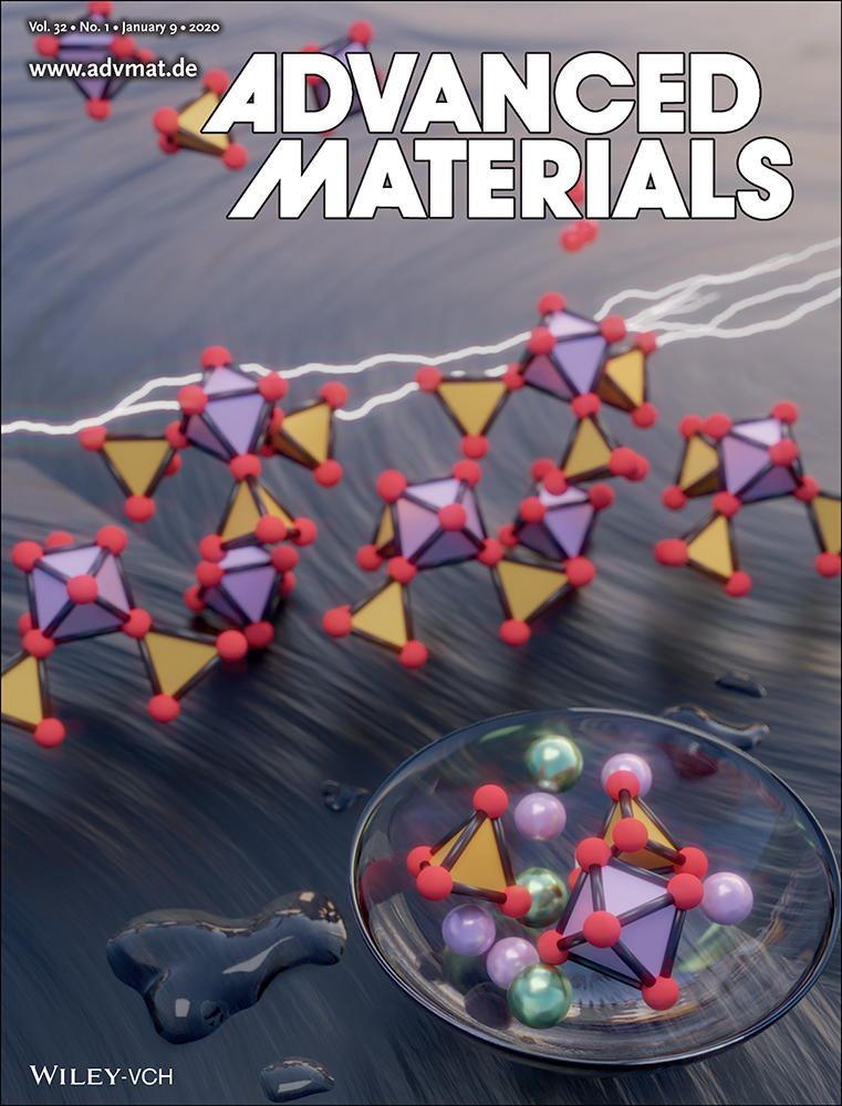
3D printable and reconfigurable liquid crystal elastomers (LCEs) that reversibly shape-morph when cycled above and below their nematic-to-isotropic transition temperature (TNI) are created, whose actuated shape can be locked-in via high-temperature UV exposure. By synthesizing LCE-based inks with light-triggerable dynamic bonds, printing can be harnessed to locally program their director alignment and UV light can be used to enable controlled network reconfiguration without requiring an imposed mechanical field. Using this integrated approach, 3D LCEs are constructed in both monolithic and heterogenous layouts that exhibit complex shape changes, and whose transformed shapes could be locked-in on demand.
3D Printing of Liquid Crystal Elastomeric Actuators with Spatially Programed Nematic Order
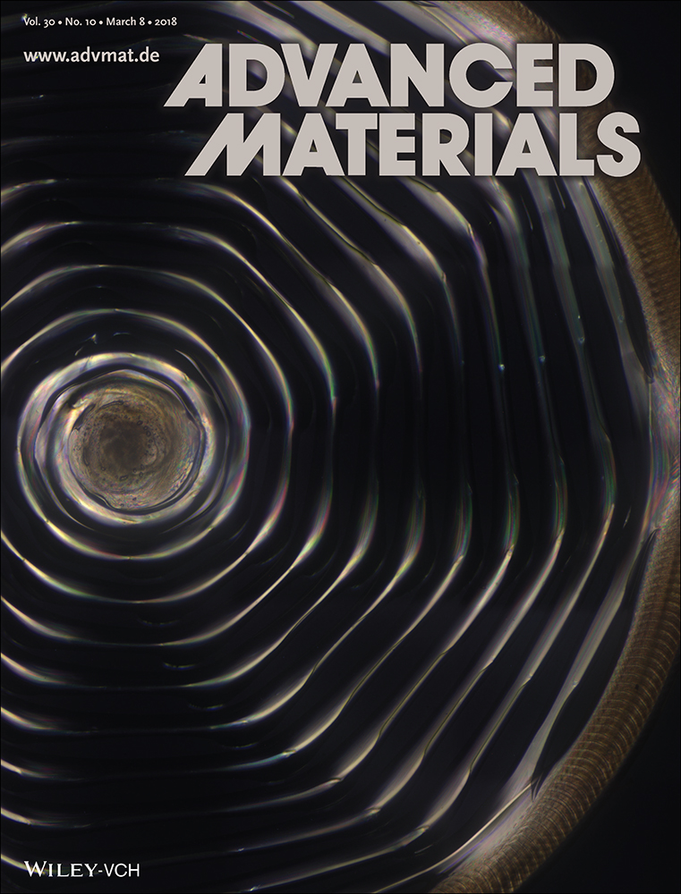
Liquid crystal elastomers (LCEs) are soft materials capable of large, reversible shape changes, which may find potential application as artificial muscles, soft robots, and dynamic functional architectures. Here, the design and additive manufacturing of LCE actuators (LCEAs) with spatially programed nematic order that exhibit large, reversible, and repeatable contraction with high specific work capacity are reported. First, a photopolymerizable, solvent-free, main-chain LCE ink is created via aza-Michael addition with the appropriate viscoelastic properties for 3D printing. Next, high operating temperature direct ink writing of LCE inks is used to align their mesogen domains along the direction of the print path. To demonstrate the power of this additive manufacturing approach, shape-morphing LCEA architectures are fabricated, which undergo reversible planar-to-3D and 3D-to-3D′ transformations on demand, that can lift significantly more weight than other LCEAs reported to date.
High-Operating-Temperature Direct Ink Writing of Mesoscale Eutectic Architectures
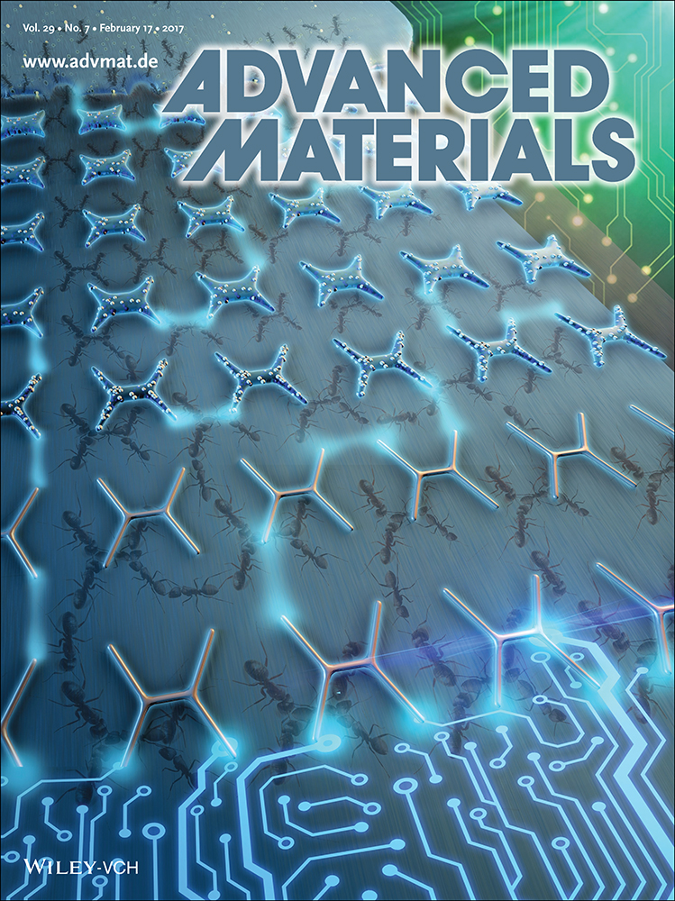
High-operating-temperature direct ink writing (HOT-DIW) of mesoscale architectures that are composed of eutectic silver chloride–potassium chloride. The molten ink undergoes directional solidification upon printing on a cold substrate. The lamellar spacing of the printed features can be varied between approximately 100 nm and 2 µm, enabling the manipulation of light in the visible and infrared range.
A combined numerical and experimental study to elucidate primary breakup dynamics in liquid metal droplet-on demand printing

Droplet-on-demand liquid metal jetting is emerging as a powerful technology for the additive manufacturing of metallic parts. The success of this method hinges on overcoming several technological challenges. The principal one among these challenges is the controlled repeatable ejection of single uniform droplets. Due to the high density and surface tension of liquid metals, the droplet ejection process occurs near the minimal extremes of the printability phase diagram, defined by acceptable ranges for the Weber (We) and Ohnesorge (Oh) numbers. In this work, we experimentally demonstrate the satellite-free ejection of pneumatically actuated molten tin droplets in this extreme corner of printability and use a combination of high-speed video analysis and volume-of-fluid modeling to elucidate the droplet dynamics. While the simulations at low Oh and We can correctly describe several aspects of the breakup process, such as an increasing tail and pinch-point near the nozzle, no single parameter set can completely capture the droplet shape at breakup. Instead, the experimental droplet dynamics appear to include features from both high and low Oh breakup. This disagreement is ascribed to the incomplete description of the droplet ejection process including wetting and exit effects near the nozzle opening and surface effects such as transient cooling and oxide formation.
Effect of carboxymethylated cellulose nanofibril concentration regime upon material forming on mechanical properties in films and filaments

It is predicted that the forest and materials from the forest will play an important role to enable the transformation from our linear present to a circular and sustainable future. Therefore, there is a need to understand the materials that can be extracted from the forest, and how to use them in an efficient manner. Here, carboxymethylated cellulose nanofibrils (CNF) from the forest are used to produce films and filaments with the aim to preserve the impressive mechanical properties of a single CNF in a macro-scale material. The mechanical properties of both the films (tensile strength of 231 MPa) and filaments (tensile strength of 645 MPa) are demonstrated to be maximized when the starting suspension is in a flowing state. This is a new insight with regards to filament spinning of CNF, and it is here argued that the three main factors contributing to the mechanical properties of the filaments are (1) the possibility to produce a self-supporting filament from a suspension, (2) the CNF alignment inside the filament and (3) the spatial homogeneity of the starting suspension. The results in this study could possibly also apply to other nanomaterials such as carbon nanotubes and silk protein fibrils, which are predicted to play a large part in future high performing applications.
Processing and reprocessing liquid crystal elastomer actuators
Liquid crystal elastomers (LCEs) have long been celebrated for their exceptional shape actuation and mechanical properties. For much of the last half century, a major focus for the field has been the development of LCE chemistries and how to process the so-called “monodomain” configurations. This foundation work has now led to a plethora of materials and processes that are enabling the demonstration of devices that are close to real-world applicability as responsive and reprocess-able actuators. In this Perspective, we review and discuss the key recent developments in the processing of actuating LCE devices. We consider how processing has been used to increase the practicality of electrical, thermal, and photo stimulation of LCE shape actuation; how dynamic chemistries are enhancing the functionality and sustainability of LCE devices; and how new additive manufacturing technologies are overcoming the processing barriers that once confined LCE actuators to thin film devices. In our outlook, we consider all these factors together and discuss what developments over the coming years will finally lead to the realization of commercial shape actuating LCE technologies.
Soft Elasticity Optimizes Dissipation in 3D-Printed Liquid Crystal Elastomers

Soft-elasticity in monodomain liquid crystal elastomers (LCEs) is promising for impact-absorbing applications where strain energy is ideally absorbed at constant stress. Conventionally, compressive and impact studies on LCEs have not been performed given the notorious difficulty synthesizing sufficiently large monodomain devices. Here, we use direct-ink writing 3D printing to fabricate bulk (>cm3) monodomain LCE devices and study their compressive soft-elasticity over 8 decades of strain rate. At quasi-static rates, the monodomain soft-elastic LCE dissipated 45% of strain energy while comparator materials dissipated less than 20%. At strain rates up to 3000 s−1, our soft-elastic monodomain LCE consistently performed closest to an ideal-impact absorber. Drop testing reveals soft-elasticity as a likely mechanism for effectively reducing the severity of impacts – with soft elastic LCEs offering a Gadd Severity Index 40% lower than a comparable isotropic elastomer. Lastly, we demonstrate tailoring deformation and buckling behavior in monodomain LCEs via the printed director orientation.
A comprehensive study of acid and base treatment of 3D printed poly (ε-caprolactone) scaffolds to tailor surface characteristics

Poly(ε-caprolactone) (PCL) chain cleavage results in the formation of polar terminal species, comprising hydroxy and carboxyl groups that enhance surface hydrophilicity and enable subsequent biofunctionalization. However, the direct effects of various acidic and basic treatments on 3D printed PCL scaffolds have not been studied from a functional perspective. In this study, we comprehensively assessed the influence of acid (hydrochloric, HCl) and base (sodium hydroxide, NaOH) catalyzed hydrolysis across different conditions on various properties of 3D printed PCL scaffolds. Analyses included testing of physiochemical and mechanical properties, and assessment of rate and stability of surface-nucleating bioactive apatite-like minerals. HCl exposure resulted in pronounced bulk degradation, as observed through limited increase in surface charge, pronounced mechanical weakening, and clear decrease in molecular weight. Conversely, NaOH treatment resulted in a more superficial degradation, with a dramatic increase in surface charge, much lower mechanical degradation, and little change in molecular weight. Indeed, the characteristics observed under acidic catalysis appeared more representative of physiological degradation. The apatite-like coating on the base-treated surface was found to be superior. Our results demonstrate the range of surface and bulk properties achievable through these accessible treatments, and support such techniques for the functionalization of PCL scaffolds for biomedical applications.
Release of lithium from 3D printed polycaprolactone scaffolds regulates macrophage and osteoclast response

The immunomodulatory effects of lithium have been reported across a range of models and contexts. Lithium appears to have a positive effect on osteogenesis in vivo, while in vitro outcomes throughout the literature are varied. Tissue engineering approaches have rarely targeted local lithium delivery within a regenerative setting. We hypothesized that part of the positive effects of lithium in vivo may be due to an immunomodulatory effect manifesting in a local environment. To achieve a sustained lithium release from scaffold constructs, we blended lithium carbonate, a soluble salt of lithium, with the biomaterial polymer polycaprolactone (PCL). We printed constructs of PCL alone, and with 5% (5Li) and 10% (10Li) lithium carbonate. Mechanical testing revealed that mechanical properties were largely retained with lithium carbonate incorporation, and we measured a consistent release of the ion over a 7 day period. The efficacy of our construct system was then assessed using a primary mouse macrophage culture, and a differentiated osteoclast culture. We found that the lithium released from constructs had a great effect on macrophage polarization, resulting in pronounced upregulation of immunomodulatory (M2) genes, and a decrease in pro-inflammatory (M1) genes. This was reflected in cytokine expression, and illustrated through immunofluorescent staining. Osteoclast activity was greatly suppressed by the lithium incorporation, with a marked effect on gene expression and actin ring formation. Our work demonstrated an effective system for local lithium delivery, confirmed the pronounced effects that lithium has on macrophage and osteoclast response, and sets the stage for further innovations in ion release for targeted tissue engineering.
Fabrication of biocompatible and bioabsorbable polycaprolactone/magnesium hydroxide 3D printed scaffolds: Degradation and in vitro osteoblasts interactions

Biodegradable polymeric 3D implants are of considerable interest for biomedical applications, however the degradation profile and bioactivity are important considerations for many clinical applications. In this context, bioresorbable magnesium hydroxide (MH) nanoparticles (NPs) (<50 nm) were blended with the degradable polymer poly (ε-caprolactone) (PCL) at concentrations of 5 and 20 wt%, and the composite was manufactured by 3D printing technology. Efficient load transfer was found between the nanofiller and matrix PCL, which was reflected in changes in the tensile properties of the MH-based composite. A statistically significant 44.3% increase in tensile modulus was achieved by the addition of 5 wt% MH, which was in agreement with the Halpin-Tsai theoretical model. The incorporation of MH in the PCL scaffolds accelerated the weight loss of the scaffolds and decreased the molecular weight of PCL over a prolonged soaking period (150 days) in PBS solution (pH 7.37, 37 ± 0.5 °C). The PCL/MH composite scaffolds were shown to be non-cytotoxic in vitro, and ion diffusion into the cell culture media promoted osteoblast metabolic activity, attachment, and proliferation, as compared to PCL-only scaffolds. Moreover, osteoblastic activity, as assessed by the expression of alkaline phosphatase, was significantly higher on the composite PCL/MH scaffold after 14 and 21 days. In summary, the 3D PCL/MH composite scaffolds could enhance osteoblastic activity and demonstrated a moderately accelerated degradation profile, which are characteristics that can be considered favorable for bone regeneration applications.
Workflow for highly porous resorbable custom 3D printed scaffolds using medical grade polymer for large volume alveolar bone regeneration

This study investigates the design, workflow, and manufacture of highly porous, resorbable additively manufactured, 3-dimensional (3D) custom scaffolds for the regeneration of large volume alveolar bone defects.
3D Bio-printed Human Skeletal Muscle Constructs for Muscle Function Restoration

A bioengineered skeletal muscle tissue as an alternative for autologous tissue flaps, which mimics the structural and functional characteristics of the native tissue, is needed for reconstructive surgery. Rapid progress in the cell-based tissue engineering principle has enabled in vitro creation of cellularized muscle-like constructs; however, the current fabrication methods are still limited to build a three-dimensional (3D) muscle construct with a highly viable, organized cellular structure with the potential for a future human trial. Here, we applied 3D bioprinting strategy to fabricate an implantable, bioengineered skeletal muscle tissue composed of human primary muscle progenitor cells (hMPCs). The bio-printed skeletal muscle tissue showed a highly organized multi-layered muscle bundle made by viable, densely packed, and aligned myofiber-like structures. Our in vivo study presented that the bio-printed muscle constructs reached 82% of functional recovery in a rodent model of tibialis anterior (TA) muscle defect at 8 weeks of post-implantation. In addition, histological and immuno-histological examinations indicated that the bio-printed muscle constructs were well integrated with host vascular and neural networks. We demonstrated the potential of the use of the 3D bio-printed skeletal muscle with a spatially organized structure that can reconstruct the extensive muscle defects.
3D Bio-printed Bio-Mask for Facial Skin Reconstruction

Skin injury to the face remains one of the greatest challenges in wound care due to the varied contours and complex movement of the face. Current treatment strategies for extensive facial burns are limited to the use of autografts, allografts, and skin substitutes, and these often result in scarring, infection, and graft failure. Development of an effective treatment modality will greatly improve the quality of life and social integration of the affected individuals. In this proof of concept study, we developed a novel strategy, called “BioMask”, which is a customized bioengineered skin substitute combined with a wound dressing layer that snugly fits onto the facial wounds. To achieve this goal, three-dimensional (3D) bioprinting principle was used to fabricate the BioMask that could be customized by patients’ clinical images such as computed tomography (CT) data. Based on a face CT image, a wound dressing material and cell-laden hydrogels were precisely dispensed and placed in a layer-by-layer fashion by the control of air pressure and 3-axis stage. The resulted miniature BioMask consisted of three layers; a porous polyurethane (PU) layer, a keratinocyte-laden hydrogel layer, and a fibroblast-laden hydrogel layer. To validate this novel concept, the bio-printed BioMask was applied to a skin wound on a pre-fabricated face-shaped structure in mice. Through this in vivo study using the 3D BioMask, skin contraction and histological examination showed the regeneration of skin tissue, consisting of epidermis and dermis layers, on the complex facial wounds. Consequently, effective and rapid restoration of aesthetic and functional facial skin would be a significant improvement to the current issues a facial wound patient experience.
3D Bio-printed Highly Elastic Hybrid Constructs for Advanced Fibrocartilaginous Tissue Regeneration

Advanced strategies to bioengineer a fibrocartilaginous tissue to restore the function of the meniscus are necessary. Currently, 3D bioprinting technologies have been employed to fabricate clinically relevant patient-specific complex constructs to address unmet clinical needs. In this study, a highly elastic hybrid construct for fibrocartilaginous regeneration is produced by co-printing a cell-laden gellan gum/fibrinogen (GG/FB) composite bio-ink together with a silk fibroin methacrylate (Sil-MA) bio-ink in an interleaved crosshatch pattern. We characterize each bio-ink formulation by measuring the rheological properties, swelling ratio, and compressive mechanical behavior. For in vitro biological evaluations, porcine primary meniscus cells (pMCs) are isolated and suspended in the GG/FB bio-ink for the printing process. The results show that the GG/FB bio-ink provides a proper cellular microenvironment for maintaining the cell viability and proliferation capacity, as well as the maturation of the pMCs in the bio-printed constructs, while the Sil-MA bio-ink offers excellent biomechanical behavior and structural integrity. More importantly, this bio-printed hybrid system shows the fibrocartilaginous tissue formation without a dimensional change in a mouse subcutaneous implantation model during the 10-week post implantation. Especially, the alignment of collagen fibers is achieved in the bio-printed hybrid constructs. The results demonstrate that this bio-printed mechanically reinforced hybrid construct offers a versatile and promising alternative for the production of advanced fibrocartilaginous tissue.
3D Bio-printed Functional and Contractile Cardiac Tissue Constructs

Bioengineering of a functional cardiac tissue composed of primary cardiomyocytes has great potential for myocardial regeneration and in vitro tissue modeling. However, its applications remain limited because the cardiac tissue is a highly organized structure with unique physiologic, biomechanical, and electrical properties. In this study, we undertook a proof-of-concept study to develop a contractile cardiac tissue with cellular organization, uniformity, and scalability by using three-dimensional (3D) bioprinting strategy. Primary cardiomyocytes were isolated from infant rat hearts and suspended in a fibrin-based bio-ink to determine the printing capability for cardiac tissue engineering. This cell-laden hydrogel was sequentially printed with a sacrificial hydrogel and a supporting polymeric frame through a 300-µm nozzle by pressured air. Bio-printed cardiac tissue constructs had a spontaneous synchronous contraction in culture, implying in vitro cardiac tissue development and maturation. Progressive cardiac tissue development was confirmed by immunostaining for α-actinin and connexin 43, indicating that cardiac tissues were formed with uniformly aligned, dense, and electromechanically coupled cardiac cells. These constructs exhibited physiologic responses to known cardiac drugs regarding beating frequency and contraction forces. In addition, Notch signaling blockade significantly accelerated development and maturation of bio-printed cardiac tissues. Our results demonstrated the feasibility of bioprinting functional cardiac tissues that could be used for tissue engineering applications and pharmaceutical purposes.

